Fluorescence In Situ Hybridization (FISH)
Definition
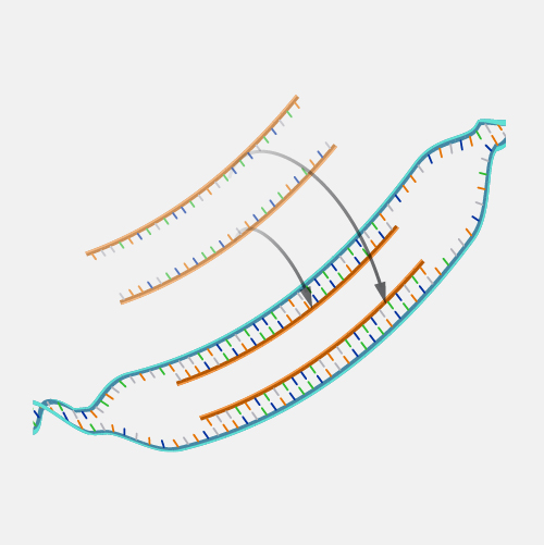
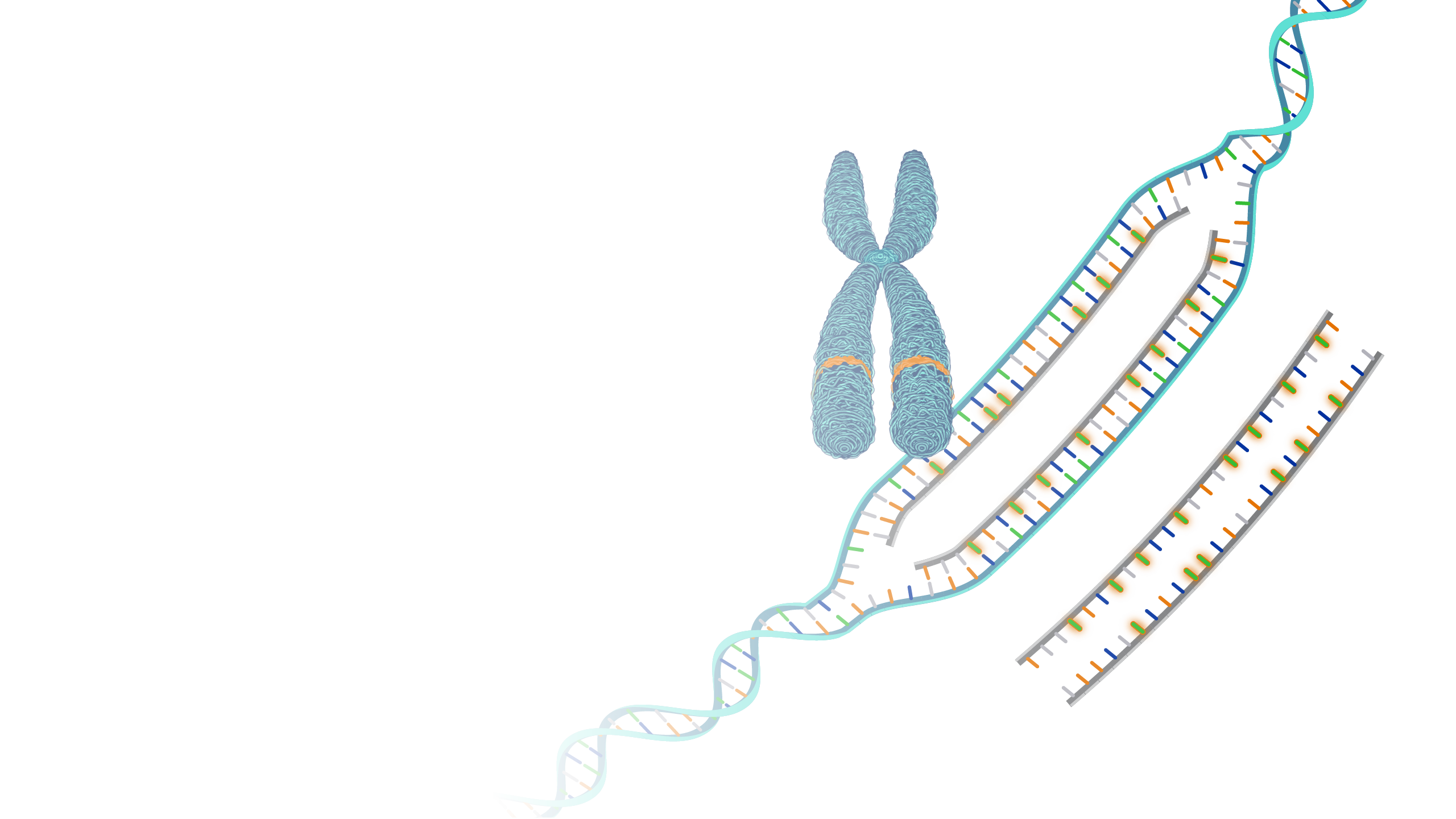
Fluorescence in situ hybridization (abbreviated FISH) is a laboratory technique used to detect and locate a specific DNA sequence on a chromosome. In this technique, the full set of chromosomes from an individual is affixed to a glass slide and then exposed to a “probe”—a small piece of purified DNA tagged with a fluorescent dye. The fluorescently labeled probe finds and then binds to its matching sequence within the set of chromosomes. With the use of a special microscope, the chromosome and sub-chromosomal location where the fluorescent probe bound can be seen.

Narration
Fluorescence in Situ Hybridization (FISH). Fluorescence in situ hybridization (FISH) is a molecular cytogenetic technique that allows the localization of a specific DNA sequence or an entire chromosome in a cell. It is utilized to diagnose genetic diseases, gene mapping, and identification of chromosomal abnormalities, and may also be used to study comparisons among the chromosomes' arrangements of genes of related species. FISH involves unwinding of the double helix structure and binding of the DNA of all probes attached to a fluorescent molecule with a specific sequence of sample DNA, which can be visualized under the fluorescent microscope.


